The Silent Swell that Spoke Cancer
1) Diagnosis
- Primary: Squamous Cell Carcinoma (SCC) of the Left Buccal Mucosa (T2N1M0, Stage III – AJCC 8th Edition).
- Secondary: Anemia of chronic disease, tobacco-induced leukoplakia changes.
2) History of Present Illness (HPI)
- Chief Complaint: Non-healing ulcer in left buccal mucosa × 3 months.
- Progression: Initially small, painless ulcer → gradually increased in size with pain on chewing & mild trismus.
- Associated Symptoms: Weight loss (4 kg in 2 months), halitosis.
- Negative History: No bleeding from ulcer, no dysphagia, no hoarseness.
3) Past Medical History
- HTN (well controlled on Amlodipine 5 mg OD).
- No prior surgeries/ICU admissions.
- Medications: Amlodipine 5 mg (Ca²⁺ channel blocker, vasodilator → lowers BP).
4) Physical Examination
- General: Cachectic, mild pallor, ECOG performance status 1.
- Oral Exam:
- Ulcer proliferative lesion, left buccal mucosa, ~3 × 2.5 cm.
- Indurated margins, non-tender, limited mobility.
- Trismus: mouth opening 2.5 cm.
- Neck: Single palpable ipsilateral cervical lymph node (1.5 cm, firm, mobile, non-tender).
- Systemic Exam: No organomegaly, chest clear, CVS normal.
5) Vitals on Admission
- HR: 92
- bpm BP: 138/84 mmHg
- RR: 18/min
- Temp: 37.2°C
- SpO₂: 97% RA
- GCS: 15/15
6) Investigations
- Baseline (Day 0):
- CBC: Hb 10.8 g/dL (mild anemia), WBC 8,500, Plt 2.1 lakh.
- RFT: Normal.
- LFT: Mildly ↑ SGOT/SGPT.
- Electrolytes: Normal.
- FNAC of cervical node: Positive for metastatic SCC.
- Biopsy (from buccal lesion): Moderately differentiated SCC.
- CXR: Normal (no lung mets).
- CECT Face & Neck: Tumor confined to left buccal mucosa, extension to retromolar trigone, ipsilateral LN involvement, no mandibular bone invasion.
7) Imaging
- CT scan: Stage III (T2N1M0).
- Chest X-ray/USG Abdomen: No distant metastasis.
8) Emergency Stabilization (Day 0 – ER)
- Symptoms: Painful non-healing ulcer in left buccal mucosa × 3 months, mild trismus, weight loss, halitosis.
- Exam: Ulcer 3 × 2.5 cm, indurated margins, palpable ipsilateral cervical LN (1.5 cm).
- Vitals: HR 92, BP 138/84, RR 18, Temp 37.2°C, SpO₂ 97%, GCS 15.
- Investigations:
- CBC: Hb 10.8 g/dL, WBC 8,500, Plt 2.1 lakh.
- RFT: Cr 0.9, Urea 24.
- LFT: Mild ↑ SGOT/SGPT.
- CECT Face & Neck: Tumor confined to left buccal mucosa + ipsilateral LN, no bone invasion.
- Biopsy: Moderately differentiated SCC.
- Treatment:
- IV fluids (DNS 100 mL/hr).
- Analgesia: Inj Tramadol 50 mg IV TDS.
- Oral hygiene: Betadine gargle QID.
- Nutrition: High-protein NG tube feed.
- Plan: Optimize for surgery
9) Day-by-Day ICU/Pre-op & Operative Course
- Day 1 – Preoperative Optimization
- Findings: Mild pallor, ECOG 1, trismus persists.
- Investigations: ECG – Normal sinus rhythm. CXR – clear lungs.
- Treatment:
- Inj Iron Sucrose 200 mg IV (to correct anemia).
- Multivitamins + high-protein diet via NG tube.
- BP controlled on Amlodipine 5 mg OD.
- Event: Pre-anesthesia clearance obtained
- Day 2 – Surgery Day
- Procedure: Wide local excision of buccal mucosa lesion + Marginal Mandibulectomy + Ipsilateral Modified Radical Neck Dissection (Levels I–IV).
- Reconstruction: Pectoralis Major Myocutaneous (PMMC) flap.
- Intra-Op Events:
- Blood loss ~500 mL (1 PRBC transfused).
- Margins taken – frozen section negative.
- Post-Op Orders:
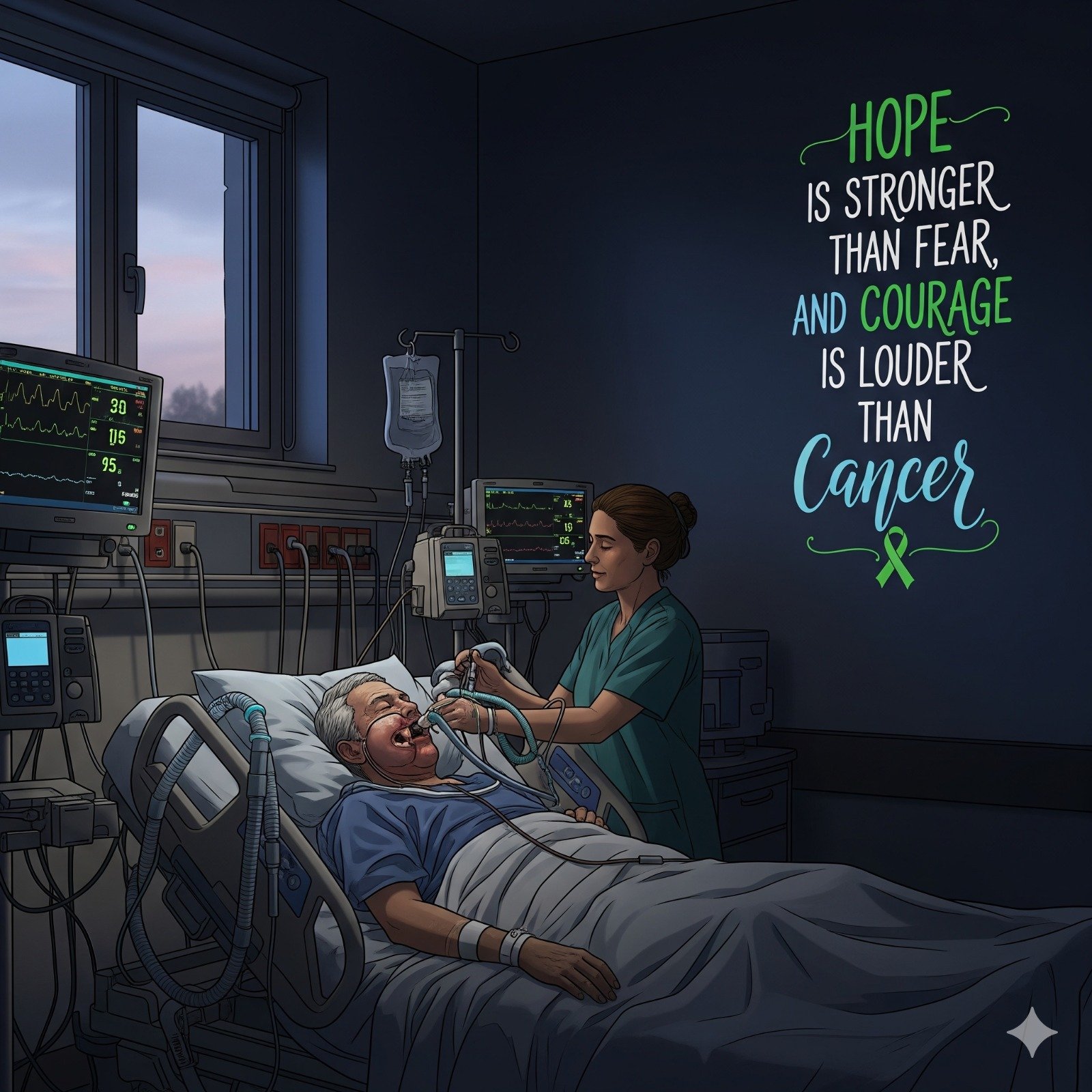
- Ventilated overnight in ICU.
- IV Ceftriaxone 1 g BD + Inj Metronidazole 500 mg TDS.
- Analgesia: PCA Morphine (1 mg bolus, lockout 10 min, max 6 mg/hr).
- IV Fluids: DNS + RL, 100 mL/hr each.
- NG feeding deferred until bowel sounds return.
- Vitals stable.
- Day 3 – Immediate Post-Op
- Event: Extubated successfully in morning.
- Findings: Flap viable (pink, warm, good capillary refill).
- Investigations: Hb 10.4 g/dL, WBC 9,200, Cr 1.0.
- Treatment:
- Restarted NG tube feeding (liquid diet, 200 mL every 3 hr).
- Inj Pantoprazole 40 mg IV OD.
- DVT prophylaxis: Enoxaparin 40 mg SC OD.
- Analgesia shifted to Inj Paracetamol 1 g IV TDS + Tramadol SOS.
- Day 4–5 – Early Recovery
- Symptoms: Pain reduced, able to sit up, no respiratory distress.
- Exam: Flap healthy, sutures intact, drains minimal serosanguinous.
- Investigations: CBC stable, electrolytes normal.
- Treatment:
- Continue NG feeding.
- Daily saline + betadine oral care.
- Physiotherapy for neck/shoulder started.
- Plan: Monitor until final histopathology report (HPR).
- Day 6–7 – Post-Op Ward Stay
- Event: Drain removed (output <30 mL/24 hr).
- HPR: Moderately differentiated SCC, clear surgical margins, 1/14 nodes positive → Stage III, T2N1M0.
- Plan: Adjuvant concurrent chemo-radiotherapy.
- Day 8 – Discharge
- Medications:
- Tab Amlodipine 5 mg OD.
- Tab Tramadol 50 mg SOS.
- Tab Pantoprazole 40 mg OD.
- Oral multivitamins, zinc.
- Advice: Oral hygiene, NG feeding until adequate mouth opening, follow-up in 2 weeks.
- Medications:
- Day 14 – OPD Follow-Up
- Findings: Flap well-settled, no infection. Mouth opening improved.
- Event: Planned for chemoradiation start.
10) Adjuvant Therapy Course
- Week 3 (Day 21) – Start of Chemoradiation
- Radiotherapy: IMRT 60 Gy in 30 fractions (2 Gy × 5 days/week).
- Chemo: Inj Cisplatin 100 mg/m² IV Day 1, 22, 43.
- Prehydration: 2 L NS + KCl + MgSO₄.
- Antiemetics: Inj Ondansetron 8 mg IV BD, Inj Dexamethasone 8 mg IV OD.
- Supportive:
- NG feeding continued.
- Oral care to prevent mucositis.
- Monitoring: CBC, RFT, Electrolytes twice weekly.
- Week 4–5 – During Radiation
- Symptoms: Oral mucositis Grade II, mild dysphagia, nausea.
- Investigations: Hb 10.2 g/dL, Cr 1.1, WBC 5,800.
- Treatment:
- Topical Lignocaine viscous before meals.
- Tab Paracetamol 650 mg TDS.
- IV fluids on chemo days.
- Week 7 – End of Radiation
- Event: Completed 60 Gy/30 fractions + 3 cycles Cisplatin.
- Symptoms: Oral mucositis resolving, weight loss ~2 kg.
- Treatment: Nutritional rehabilitation, high-protein feeds, antifungal (Fluconazole) for oral thrush.
11) 3-Month Follow-Up
- Findings: Healing flap, good oral intake, no recurrence clinically.
- Investigations: CECT Neck – no residual/recurrent disease.
- Patient Status: ECOG 0, returned to daily activities.
12) Key Clinical Pearls
- Early biopsy of any non-healing oral ulcer (>2 weeks) is critical.
- Tobacco + betel nut chewing = strongest risk factor.
- Surgery + adjuvant chemoradiation = best outcome in Stage III.
- Regular flap monitoring avoids catastrophic necrosis.
- Nutrition & oral hygiene play a major role in recovery.
13) Case Storytelling
“Mr. R, a 52-year-old farmer with a long history of chewing tobacco, walked in with a wound inside his cheek that refused to heal. For months, he thought it was a simple sore — until pain made even a sip of water a challenge. On the day of surgery, a team worked meticulously to remove the cancer and rebuild his cheek with muscle from his chest. A week later, as he smiled weakly and sipped his first spoon of soup, he whispered — ‘Doctor, it feels like a new life.’”
14) MCQ Q&A Section
- Q1. Most common histological type of buccal mucosa cancer?
Ans: Squamous Cell Carcinoma (≈90%). - Q2. Best adjuvant therapy for Stage III oral cavity SCC post-surgery?
Ans: Concurrent chemoradiation (Cisplatin-based + IMRT). - Q3. Radiation dose commonly used in buccal mucosa SCC?
Ans: 60–66 Gy in 30–33 fractions. - Q4. Which flap is most commonly used for reconstruction in buccal mucosa carcinoma?
Ans: Pectoralis Major Myocutaneous Flap (PMMC). - Q5. Most important prognostic factor?
Ans: Nodal status (lymph node involvement).