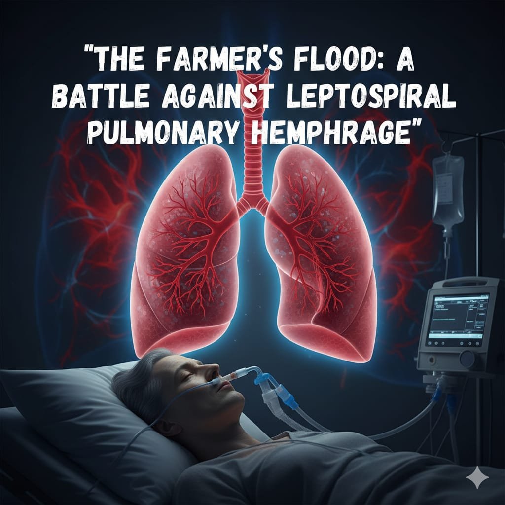
The Farmer’s Flood: A Battle Against Leptospiral Pulmonary Hemorrhage
1) Diagnosis
- Primary Diagnosis: Severe Leptospirosis with Pulmonary Hemorrhage Syndrome (LPHS)
- Secondary/Complicating Conditions: Acute Kidney Injury (AKI), Septic Shock, ARDS, Thrombocytopenia
2) History of Present Illness (HPI)
- Chief complaints: Fever (7 days), Jaundice (5 days), Hemoptysis + Breathlessness (2 days)
- Progression: Initially fever with myalgia → developed jaundice → within 48 hrs developed hemoptysis and respiratory distress
- Associated symptoms: Oliguria, malaise, calf tenderness
- Negative history: No history of TB, no prior lung disease, no dengue exposure, no drug allergies
3) Past Medical History
- Diseases: No known HTN, DM, CAD
- ICU Admissions: None in past
- Medications: None regular
4) Physical Examination
- General: Ill-looking, icteric, pale, in respiratory distress
- CVS: Tachycardia, no murmur, JVP mildly raise
- RS: Bilateral crepitation’s, coarse crackles, decreased air entry (pulmonary hemorrhage).
- CNS: GCS 13/15, no focal deficit
- Abdomen: Hepatomegaly, mild ascites
5) Vitals on Admission
- HR: 124/min (tachycardia)
- BP: 84/50 mmHg (hypotension)
- RR: 34/min (tachypnea)
- Temp: 101.8°F
- SpO₂: 72% on room air
- GCS: E4V3M6 (13/15)
6) Echocardiography (ECHO)
- LV size & EF: Normal, EF 55%
- Wall motion: No regional wall motion abnormality
- Valves: Normal
- LA/RV: Normal
- Pericardial effusion: Absent
7) Investigations
Day 0 (Admission):
- CBC: Hb 10.2, WBC 18,500, Plt 28,000
- RFT: Urea 110, Creatinine 3.8
- Electrolytes: Na⁺ 132, K⁺ 5.6, Ca²⁺ 7.8, Mg²⁺ 1.5
- LFT: Bilirubin 12 (direct 8), AST 180, ALT 210
- ABG: PaO₂ 52 mmHg, PaCO₂ 32, HCO₃⁻ 18, Lactate 3.8
- BNP: 180 pg/mL (normal)
- Troponin I: Negative
- Thyroid Panel: Normal
- CRP: 148, Procalcitonin: 8.2
- Coagulation: INR 1.8, aPTT 42 sec
- Lactate: 3.8 mmol/L
- Leptospira IgM ELISA: Positive
- Dengue NS1/IgM: Negative
- Malaria smear: Negative
| Day | Na+ | K+ | Creatinine | PaO2/FiO 2 | CRP | Platelets |
| 0 | 132 | 3.8 | 3.8 | 85 | 148 | 28k |
| 1 | 134 | 4.5 | 4.5 | 72 | 162 | 20k |
| 2 | 136 | 6.2 | 5.2 | 90 | 140 | 18k |
| 3 | 138 | 5.1 | 3.9(CRRT) | 180 | 98 | 65k |
8) Imaging
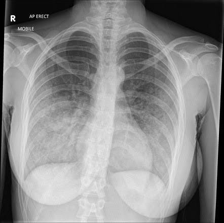
- CXR: Bilateral diffuse alveolar infiltrates (ARDS pattern)
- USG Abdomen: Hepatomegaly, mild ascites
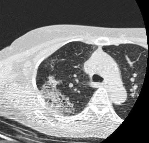
- CT Chest (Day 2): Diffuse ground-glass opacities with patchy alveolar hemorrhage
9) ECG & RBS
- ECG: Sinus tachycardia, no ischemic changes
- RBS: 122 mg/dL
10) Emergency Stabilization (Day 0 – ER)
- Airway/Breathing: O₂ via NRBM → Intubated within 2 hrs (ARDS with PaO₂ < 60)
- Circulation: 2 large-bore IVs, restricted crystalloids (60 mL/hr), Noradrenaline started @ 0.1 mcg/kg/min
- Drugs:
- IV Penicillin G 1.5 MU q6h (antimicrobial)
- IV Methylprednisolone 1 mg/kg/day (immune-mediated hemorrhage)
- IV Pantoprazole 40 mg OD (stress ulcer prophylaxis)
- Blood products: 4 units platelets (plt < 20k with bleeding)
11) Day-by-Day ICU Care
- Day 1 – ICU
- The patient was admitted to the ICU with severe hypoxemic respiratory failure and was managed on the ARDS protocol with lung-protective ventilation (tidal volume 6 mL/kg, PEEP 12 cm H₂O, FiO₂ 80%). Despite optimization, oxygenation remained borderline. Hemodynamically, the patient was unstable, requiring norepinephrine infusion titrated to maintain MAP above 65 mmHg. Renal parameters showed progressive deterioration, with creatinine rising from 3.8 mg/dL to 4.5 mg/dL accompanied by oliguria, indicating evolving acute kidney injury. Hematologically, the patient developed worsening thrombocytopenia (platelet count ~20,000/µL) and hemoptysis. This warranted transfusion support, and the patient received 2 units of packed red blood cells along with 4 units of platelets. Despite aggressive interventions, the condition remained critical with high risk of multi-organ dysfunction.
- Day 2 – ICU
- On the second ICU day, the patient remained on ARDS protocol ventilation with high PEEP support, though FiO₂ was gradually titrated down to 70% as oxygenation showed minimal improvement. Hemodynamically, the norepinephrine infusion was continued at 0.2– 0.3 µg/kg/min, maintaining MAP just above 65 mmHg. Renal dysfunction persisted with oliguria and rising urea/creatinine levels, and nephrology was consulted for possible initiation of renal replacement therapy. Hematologically, platelets remained low (~25,000/µL), necessitating one more platelet transfusion to cover risk of mucosal bleed. Hemoptysis reduced after correction of counts. Supportive measures included stress ulcer prophylaxis with IV pantoprazole, DVT prophylaxis using mechanical compression devices (pharmacologic anticoagulation was withheld due to thrombocytopenia), and broad-spectrum antibiotics were escalated considering the possibility of sepsis driving the worsening organ dysfunction.
- Day 3 – ICU
- By the third ICU day, the patient’s oxygenation began showing signs of stabilization with FiO₂ down to 60% while maintaining adequate saturations on the ARDS protocol. Hemodynamics improved, and norepinephrine was slowly tapered to 0.1 µg/kg/min. Renal function, however, worsened with creatinine climbing further, and the patient was initiated on slow low-efficiency dialysis (SLED). Platelet counts improved modestly after transfusion support, reaching 45,000/µL, and no further bleeding episodes were noted. Enteral feeding was gradually restarted via Ryle’s tube with a high-protein, renal-adjusted formula. Daily labs and ABGs were closely monitored, and meticulous input-output charting was maintained. The patient continued to require close ICU monitoring but showed early signs of stabilization.
- Day 4-6 – ICU
- Over the next 48–72 hours, the patient showed progressive recovery. FiO₂ requirements decreased to 40%, and sedation was lightened, allowing for spontaneous breathing trials. Vasopressors were successfully weaned off, and urine output improved with continued renal support. By Day 6, the patient was successfully extubated onto high-flow nasal oxygen (HFNO) with good oxygenation. Hemodynamics were stable without pressor, and renal function stabilized with intermittent dialysis. With improving platelet counts and absence of active bleeding, the risk of hemorrhage reduced significantly.
- Ward Course (Step-Down Management)
- Once shifted to the ward, the focus was on rehabilitation and supportive care. The patient continued on HFNO (40 L/min, FiO₂ 35%), chest physiotherapy, and incentive spirometry for lung recovery. Antibiotics were continued as per cultures, and dialysis was given intermittently based on renal function trends. Nutrition was optimized with a renal protective, high-calorie diet, and physiotherapy was intensified to regain muscle strength lost during prolonged ICU stay. Serial labs, including CBC, renal profile, LFT, and coagulation profile, were monitored. Over the course of 10 days in the ward, the patient achieved steady improvement, with renal parameters slowly stabilizing and respiratory status improving, eventually allowing weaning to room air.
12) Discharge Medicines
- Doxycycline 100 mg BD × 7 days (eradication)
- Tab Pantoprazole 40 mg OD (GI protection)
- Multivitamin + High-protein diet
- No anticoagulation (risk of bleeding)
13) Key Notes / Clinical Pearls
- Leptospira can mimic dengue/malaria/hepatitis – always suspect in flood season.
- Weil’s triad: Jaundice + AKI + Hemorrhage = Severe leptospirosis.
- Pulmonary hemorrhage → treat with early ventilation, steroids, transfusions.
- CRRT is lifesaving in AKI + sepsis.
- Antibiotics (Penicillin/Ceftriaxone) must be started early.
14) Case Storytelling
“A 32-year-old farmer returned from his flooded fields with fever and jaundice. Within days, he began coughing blood, drowning in his own lungs. In ICU, his kidneys shut down, his lungs bled, and his heart struggled. Yet, with timely ventilation, dialysis, antibiotics, and transfusions — he fought back. After 12 days, he walked out alive, one of the rare survivors of Leptospiral Pulmonary Hemorrhage Syndrome.”
15) MCQ Q&A Section (Learning Pearls)
- Q1: What triad defines Weil’s disease?
Ans: Jaundice + AKI + Hemorrhage - Q2: Which antibiotic is first-line in severe leptospirosis?
Ans: IV Penicillin G (1.5 MU q6h) or Ceftriaxone 2 g q12h - Q3: Why avoid aggressive fluids in LPHS?
Ans: It worsens pulmonary hemorrhage and ARDS - Q4: What ICU strategy improves oxygenation in ARDS?
Ans: Low tidal volume ventilation, high PEEP, prone positioning - Q5: What marker differentiates bacterial vs sterile inflammation?
Ans: Procalcitonin (PCT)